
Colorectal cancer is very common and affects both men and women equally. Treatment is currently multi-systemic. This means that a range of treatments are applied—chemotherapy, surgery, and radiotherapy. The treatment results from collaboration among specialists in the group called the Oncology Council or Multi-Disciplinary Team (MDT).
HEALING WITH HOPE
Detection of metastases
A CT scan of the abdomen and chest is necessary for staging. If there are no findings, the disease is considered localized and more curable. If there are secondary locations in the liver, lungs, etc., then a systemic treatment strategy is needed as a first step. The examination must be done intravenously and orally with contrast, which requires good kidney function.
Rectal-pelvic magnetic resonance imaging (MRI) has played a central role in the local staging of rectal cancer. An important role for this was played by the work of the imaging radiologist Dr. Gina Brown from London's Royal Marsden Hospital, who set the standards for assessing and staging the depth of tumor penetration, the "purity" of the peripheral resection boundary (CRM), the presence of vascular tumors (LMVI), and lymph nodes. Proper MRI evaluation is essential in selecting patients who require preoperative chemo-radiotherapy.
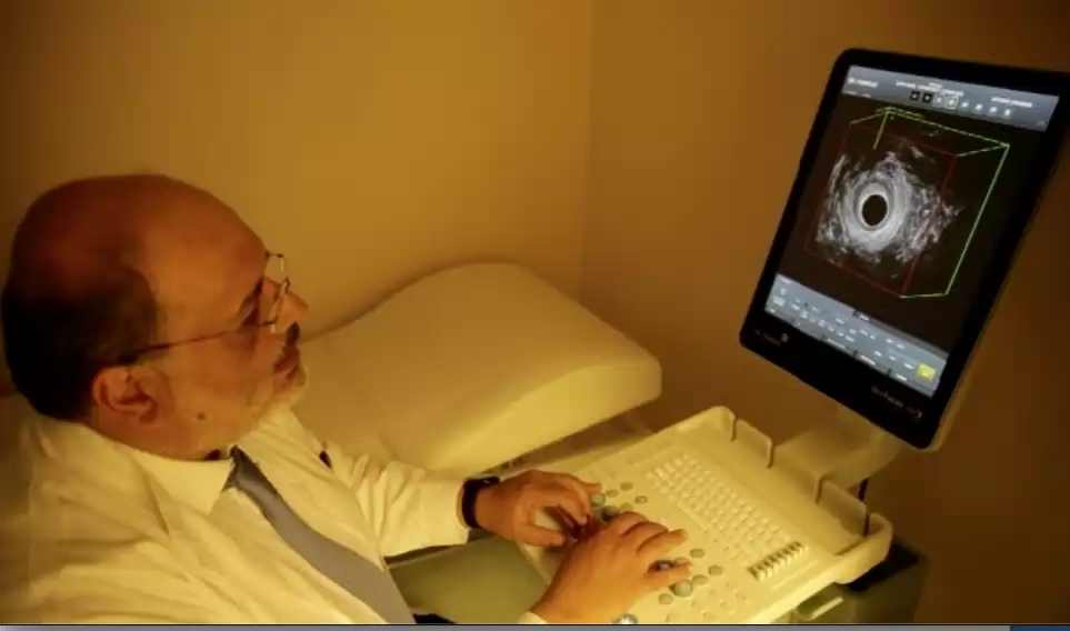
Essential for tumor selection for endoscopic resection. Intrarectal ultrasound is a valuable tool for staging polyps and rectal tumors. The 360-degree rotating head with high frequencies of 10-16 MHz can distinguish 5 layers of the rectal wall and see if the tumor is early or extending into fat. Regional lymph nodes are also imaging. EUS has greater accuracy than MRI in early tumors, which is why it is part of the protocol by which early tumors are selected, which can be excluded with TEMS or the GRECARR protocol.
PET SCAN CT is performed in special cases when there is a need to check for any micro-metastases that are not imaged on conventional CT scan. It is often performed when elevated cancer markers are found that raise suspicion of spread.
For nearly 2 centuries now, Surgery has been considered the main treatment for rectal cancer. Although this is still true, in recent years radiotherapy and drug therapy have brought significant changes to indications as well as surgical techniques. Treatment regimens are constantly evolving, and most importantly, treatment is beginning and becoming more and more individualized, always within the framework of protocols. Communication among the various specialties involved in treatment takes place through the Multidisciplinary Team, that is, the interdisciplinary council of specialists. All patients diagnosed with rectal cancer must follow coordinated protocols from surgeons, oncologists, radiotherapists, gastroenterologists, radiologists, and others. A holistic approach to the patient and psychological support are also important. And it is extremely important not to forget that in cancer treatment, survival is not only important, but quality of life is equally important. No treatment is applied unless its impact on the patient's quality of life is also examined.
The rectum is surrounded by a cytolipotic tissue filled with lymph nodes called the MESORECTUM. Radical excision of rectal cancer requires the removal of this tissue and is called TOTAL MESORECTAL EXCISION (TOTAL MESORECTAL EXCISION). This was established by the British surgeon shown BILL HEALD. MEI can be performed either openly or laparoscopic/robotic – there is no difference in oncological effectiveness between the various methods. MEI achieves cancer treatment with very low rates of local recurrence.
TEMS – Transanal Endoscopic Microsurgery is a transrectal (through the rectum) endoscopic surgery that removes rectal tumors without the need for abdominal incisions and without laparoscopy. TEMS is performed under general anesthesia and requires bowel preparation as in colonoscopy. The surgical endoscope is inserted into the rectum, and the surgery is performed with the help of an endoscopic camera and laparoscopic instruments entirely from the rectum. The TEMS technique is not the same as colonoscopy; the tumor or polyp is completely removed with all layers of the intestinal wall, and the resulting loss is closed with endoscopic sutures. Recovery is very rapid. Hospitalization usually lasts only one night. There is no significant postoperative pain, and most patients do not need any pain relief at all. The risk of complications is very low, almost as high as in colonoscopy. TEMS was invented in 1980 by the German shown GERHARD BUESS, from whom I was fortunate to train. I am a TEMS specialist and have performed over 400 TEMS surgeries at King's College Hospital and in Athens.
Laparoscopic Surgery has been an established technique for 20 years for all abdominal surgeries. Laparoscopic surgery for intestinal diseases such as cancer, inflammatory disease, diverticulitis, and others has many advantages, which have been proven by numerous research studies showing that it is safe, has faster recovery, fewer complications, and better oncological outcomes.
The smart and effective method of TRANSANAL TOTAL MESORECTAL EXCISION - TaTME is a combination of laparoscopic TEMS and transrectal TEMS and combines the advantages of both. The first publication was made in 2009 by the pictured Dr. Patricia Sylla et al from Massachusetts General Hospital, Boston. TaTME allows the sphincter to be maintained in very low rectal volumes. I have been applying TaTME since 2013 to volumes of the lower and middle third of the rectum.
Robotic surgery is a variation of laparoscopic surgery with the addition of a robot that controls three arms controlled by the surgeon via a console. In reality, it is not a true robot, since the machine does not operate autonomously but is a system that offers stability and precision of movement. There are many ongoing studies showing so far that robotic surgery is equivalent to laparoscopic surgery for colon cancer without any additional advantage. It is possible that in the future, with advances in robotic technology, the advantages will increase, but for now it seems to add more anesthesia time and financial cost, without any other clear advantage.
The end of surgery and other treatment is the beginning of the road to recovery and the path to healing. The journey is long, taking about 5 years, during which there must be monitoring and testing, the so-called follow-up. There must be a balance between the tests performed to detect any recurrence early with the aim of treatment, on the one hand. On the other hand, every person must be allowed to live as normal a life as possible without frequent visits and tests affecting their quality of life. At King's College Hospital, we developed a follow-up scheme capable of detecting relapses at a treatable stage so that overall survival exceeds 80%, and moreover, without affecting quality of life. I apply this protocol in Athens as well.
In the past, 30-40% of patients with rectal Ca ended up with a colostomy (unnatural location). Today, fortunately, this percentage is below 10% in the hands of specialist rectal surgeons. A terminal temporary Hartmann colostomy may only be needed in cases of emergency surgery for perforation or intestinal obstruction. New anatomy techniques, transsphincter resection, TEMS, and radiotherapy have reduced the need for ventricular perineal resection.
In many cases of "low rectum excision with total resection of the mediastorectum," a temporary bypass of the anastomosis is necessary. This is more necessary in low anastomoses. The reason is that the lower rectum does not have good perfusion. Good perfusion is necessary to heal the anastomosis (connection). If the healing of the anastomosis is incomplete, a serious complication occurs: anastomotic escape. The complication is avoided by creating a temporary ileostomy, that is, bypassing the anastomosis by externalizing a small intestine helix. The ileostomy produces a green bile fluid. It needs to remain until the anastomosis is controlled and intact. A temporary ileostomy can be closed in 3 weeks, but in practice, it lasts about 3 months for various reasons. After the ileostomy is closed, the rectum works normally again, but an adjustment period may be needed (see below).
The first priority is always the radical elimination of the cancer. The second priority is the preservation of the clamps and their normal function. The specialty of Colorectal Surgery has been developed for this purpose, and the ways to preserve the sphincters are: a) Transanal TME - Transrective TME with pull-through anastomosis b) Transfilarative excision and colo-anal anastomosis c) TEMS after radiotherapy on tumors that are suitable d) Application of the Watch & Wait protocol with a close follow-up. The patient should be aware that very low anastomes may not be perfectly functional. "Low Resection Syndrome" - LAR may develop, with frequent bowel movements and straining. LAR is treated with pelvic floor physiotherapy and usually improves within a year.
Radiotherapy has become one of the main treatments in rectal cancer since the 1990s, and its role is continuously increasing. Radiotherapy, regardless of protocol, offers the following: a) reduction of local recurrence b) increase overall survival c) increase in free disease survival. Radiotherapy can be combined in 4 forms: 1) Postoperative radiotherapy 2) Preoperative radiotherapy short protocol 3) Preoperative radiotherapy long protocol 4) Radiotherapy in special local excision protocols.
Postoperative radiotherapy is applied when the tumor appears to be relatively early-stage and fully surgically exclusible, but in the pathological preparation it is found to have infiltrated the lymph nodes. In this case, radiotherapy combined with mild chemotherapy with pills (capectabine) reduces the risk of local recurrence.
The short preoperative radiotherapy regimen was established by the Swedes. It includes radiotherapy for 5 consecutive days with 25 Gy and surgery 5 days later. Swedish and Dutch studies have shown that this strategy reduces local recurrence and appears to increase survival as well. The short regimen is not often applied except for orthosigmoid tumors or for specific indications.
The long radiotherapy regimen has roughly double the dose (50 Gy) and is usually given in 25-28 doses lasting up to 6-10 weeks. Surgery is performed 6 weeks to 3 months after the end of radiotherapy because the treatment continues to work after the sessions end. The short and long regimens are equivalent in terms of local recurrence and survival, but the long regimen has the following advantages. 1) In 20% of patients, complete tumor remission can be achieved, so these patients may not undergo surgery (enter the WATCH & WAIT protocol) or undergo local TEMS resection according to the GRECARR 2 study. 2) In tumors that are locally extensive (LARC - Locally Advanced Rectal Cancer) and at risk of surgical CRM resection, the long-term radiotherapy regimen can lower the tumor stage ("Downstaging") and allow removal with "clear" limits. Today, the majority of patients undergo a long-term preoperative radiotherapy regimen.
We now know that local excision after radiotherapy is feasible even in stages larger than Stage I when the patients and tumors being treated are selected based on scientific criteria. There are many studies on the subject, the most recent from France and the team of the pioneering Professor Roullier, with whom I was fortunate to train. The study is GRECARR 2, and its results were published in 2020. What does the study tell us? That in selected tumors of stage T2 and T3 we can
a) to implement radiotherapy and chemotherapy
b) For tumors that show shrinkage, local endoscopic excision should be performed.
Following patients for 5 years, no difference was found in either local recurrence or five-year survival. Tumor selection must be made with strict criteria; also, if there is no initial good response to radiotherapy, radical surgery may ultimately become necessary. However, this study, funded by the National Cancer Center of France, carries an important message of hope—that for a large segment of patients with rectal cancer, combined therapy with radiochemotherapy and endoscopic TEMS / TAMIS surgery is now a first-line treatment and must be offered.

In recent years, a pioneering procedure has been established, first applied by the famous Brazilian oncologist Angelita Habr Gama (Photo). Habr Gama was the first to dare to argue that some rectal cancers can be treated without surgery but only with radiation therapy and chemotherapy. She proved this through work on thousands of patients and is now accepted and applied throughout Europe. Of course, this Watch and Wait protocol is not for all cancers but for those lucky ones, who make up about 15-20% and who, after radiation therapy and chemotherapy, have a complete clinical response. In these cases, instead of proceeding immediately with surgery, we wait with monitoring, and if the tumor recurs, we operate. If it does not recur, these patients never make it to surgery.
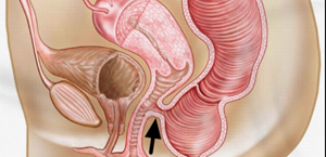
What we call "cancer" is adenocarcinoma originating from the epithelium of the intestinal lumen. There are other tumors originating from other parts of the intestinal tissue, with different progression and requiring different treatment. These tumors are GIST (formerly called sarcomas), NET - Neuroendocrine tumors (also called carcinoids), Lymphomas, Melanomas, etc.
Carcinoid tumors are relatively benign but can metastasize. Their broader name is Neuroendocrine Tumors - Neuroendocrine Tumors - NET, and they can also develop in the pancreas and other organs. In some cases, carcinoid tumors produce hormones and cause Carcinoid Syndrome, characterized by vasomotor phenomena, fluctuations in blood pressure, vasodilation, diarrhea, abdominal pain, etc.
Diagnosis is made through routine tomography scans as well as hormone detection through blood tests such as Chromogranin A, determination of catabolism derivatives in urine 5-HIAA, Octreoscan scintigraphy, etc. Treatment is mainly surgical. Small rectal carcinoids can be excluded with endoscopic local TEMS resection.
These tumors were formerly called paleomysarcomas, but today they are called stromal tumors, GASTROINTESTINAL STROMAL TUMOURS - GIST. Today, we believe they originate from the Cajal cells of the intestine. Treatment is mainly surgical. An important part of the treatment is the administration of the drug IMATINIB. This drug blocks an oncoprotein produced by the KIT oncogene and has a dramatic effect on GIST tumors.
