The pelvic floor is the muscles and ligaments that support the bladder, uterus, and rectum. When they relax or don't coordinate, incontinence, prolapse, or a feeling that bowel movements don't end.
The pelvic cavity contains important organs and is affected by many pathologies affecting multiple functions.
The pelvis is divided into three compartments: the anterior (which contains the bladder/urethra), the medial (which contains the uterus/vagina in the woman and the prostate in the man), and the posterior (which contains the rectum).
Symptoms of pelvic diseases may start in one compartment/organ, but most often extend to at least one additional compartment. Also, any therapeutic or surgical intervention in one compartment has effects (whether beneficial or side effects) on the other compartments. Therefore, a comprehensive approach to the problem is essential, and coordination among specialists—the rectal surgeon, gynecologist, urologist, physiotherapist—must design a joint treatment plan.
The pelvic floor is a funnel-shaped surface that attaches around the pelvic bones. It has two openings, the urogenital foramen and the rectal foramen, and between these two openings lies the body of the perineum. The pelvic floor provides the following functions:
● Support of the abdominal viscera
● Providing resistance and support for activities such as weightlifting, coughing, etc.
● Urine and fecal continence
It consists of a network of strong muscles that support each other, and their combined action provides the power necessary for chastity, as well as the coordinated action required for urination, defecation, and sexual function.
Sphincter incontinence followed by stool loss is a symptom that can be caused by many conditions. Unfortunately, it is very common. Also, unfortunately, many sufferers suffer silently without consulting a doctor, except when the condition reaches an extreme level. This is due to the natural reluctance to discuss a personal symptom even with doctors, as well as many patients' impression that it is a condition for which not much can be done—which is, of course, wrong. On the contrary, delayed treatment leads to muscle atrophy, the development of autonomic neuropathy, and progression of pelvic floor relaxation with age, making treatment more difficult.
Incontinence is a symptom that affects 3-5% of people. It's a symptom not everyone feels comfortable discussing, and even seeking help from a doctor is often postponed for a long time in the hope that the condition will be temporary. It can be urinary incontinence or fecal incontinence, or, in 30-40% of cases, a combination of both.
Incontinence is the inability to control the sphincters, resulting in loss of gas, or feces.
The anal sphincters are the internal sphincter, responsible for the continuous, involuntary contraction that creates the tone of the sphincter or the 'resting pressure,' and the external sphincter, responsible for the voluntary contraction that suppresses the emptying during the epithesis period . There are also the lift muscles and other pelvic floor muscles that help control incontinence, especially if there is sphincter trauma.
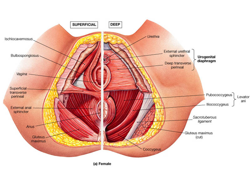
Sphincter problem, mainly damage from injury. The most common injury is childbirth in women, but there are other types of injuries, either accidents or sexual activity. Finally, there is also iatrogenic injury after surgeries. This can be... intentional, as in lateral sphincterotomy or ring dilation for fissures. This rather dangerous surgical technique is something we all learned at some point but should no longer be used under any circumstances. Injuries during childbirth are very common and are generally repaired by obstetricians with good short-term results. However, in some cases, incontinence begins to manifest either immediately, months or even years later. It is not uncommon for women to have incontinence 20-30 years after their last childbirth and to find large sphincter deficiencies on the ultrasound, which they have lived with all these years. The explanation is that at a young age, all the muscles in the body are vigorous, and the lifts and other pelvic floor muscles manage to compensate for the poor function of the sphincters for a long time. However, with advanced age, they begin to weaken and incontinence sets in. It is then difficult to correct the problem surgically because the muscles have remained denervated and atrophic for so long that the best solution is neuromodulatory therapy.
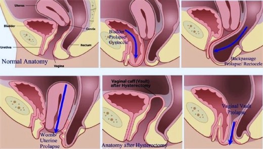
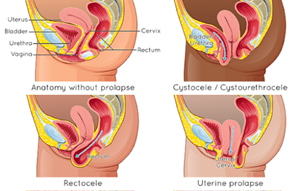
In many women, the rectum widens and causes a protrusion inside the vagina. This is called rectocele and is a common problem, especially in people over 40 and if there has been previous labor.
A rectocele causes a lump inside the vagina with a feeling of heaviness and discomfort. What many patients don't realize is that the rectocele may be responsible for a defecation disorder. The contents of the rectum pool during defecation in the rectocele instead of being pushed outward. This creates a feeling of incomplete bowel movement and the need to go to the bathroom again shortly after bowel movements. Additionally, it causes stool leakage when it is watery. This type of incontinence is revealed in tests a) defectomy, b) magnetic proctography.
Internal rectal prolapse (insertion) causes incomplete rectal emptying, pressure for repeated frequent bowel movements, and incontinence when watery stools are present. Prolapse has 5 degrees according to the OXFORD classification; the first 4 degrees are internal prolapse, and the 5th external prolapse of total fat. Only 5th-degree prolapse is operated on directly; the remaining 4 categories must first receive conservative treatment - biofeedback, and if it fails, surgery is then discussed. Additionally, chronic pressure from the prolaptic rectum causes neuropathy of the obedient nerves that contributes to incontinence. You can read more about this problem on the page on rectal prolapse.
Every neuropathy can affect the function of the sphincters. Examples include diabetic neuropathy, multiple sclerosis, etc. Also, chronic constipation and multiple pregnancies can cause neuropathy of the sphincter nerves. The consequence is a disorder of sphincter function. Damage to the sacral nerves from injuries or tumors of the spine cause combined urinary and fecal incontinence. In acute and severe cases, this takes the form of horseshoe syndrome (Cauda Equina), but in other cases the manifestation is gradual and more difficult to diagnose. The function of the sphincters and nerves is investigated by manometry—rectal physiology, electromyogram, and neurography of the vulgar nerves.
Diagnostic testing for incontinence must be thorough because treatment depends entirely on the correct diagnosis of the cause of the problem.
Incontinence is not a condition; it is a symptom. The cause of the symptom must always be found.
Diagnostic testing includes:
1. Clinical evaluation - Proctoscopy
2. Scoring
3. Three-dimensional intrarectal ultrasound
4. Manometry - rectal physiology
5. Decompression
6. Magnetic dynamic proctography
7. Endoscopic evaluation
Normal hemorrhoids are naturally designed to provide anal sealing, thus protecting against incontinence. However, if there is hemorrhoiditis and protruding grade 4 hemorrhoids, then mucus secretion creates a feeling of incontinence. In reality, it is mucus and not typical incontinence. An intra-rectal ultrasound should always be performed to rule out sphincter deficiency, and if the sphincters are intact, then hemorrhoidectomy solves the problem.
It involves taking a medical history, clinical examination, and a proctoscopy at the clinic. This determines the possible clinical cause based on the history, especially the obstetric history or previous surgeries in the area, presence of diabetes and other neuropathy causes, etc. Examination of the perineum reveals possible scarring, relaxation, or fistulas. Digital examination and proctoscopy reveal the tone of the sphincters and pelvic floor, as well as the presence of other anal causes of incontinence.
For the best methodological approach to assessing incontinence, there are various scores that "score" the severity of the specific case. This is done through special questionnaires that check bowel lierity and quality of life. The score the patient collects helps in making treatment decisions as well as in predicting the outcome.
The three-dimensional endorectal ultrasound - 3D Endorectal Ultrasound - is a fast and painless method for imaging the rectum and anal canal with high accuracy in all pathologies in the area. In incontinence, it is valuable for the accurate imaging of the sphincters and pelvic floor, and no decision can be made without the evaluation of the ultrasound image. Savvas Papagrigoriadis is certified in intrarectal ultrasound by the American Society of Colon and Rectal Surgeons with 20 years of experience in its use.
When sphincteral incontinence is present, a necessary test is rectal manometry – physiology. During the examination, a thin multichannel catheter with various sensors and a balloon is inserted into the rectum after an enema. The catheter is connected to an electronic processor and a computer. The patient executes a series of commands, and the device records a variety of data from the contraction/function of the muscles/sphincters, as well as measurements of the sensory sensitivity of the anal canal, the presence of reflexes, and the presence of incontinence. The findings correlate with the imaging seen in the three-dimensional endorectal ultrasound and other imaging tests. A manometry test can show if there is dysfunction of the medial sphincter, dysfunction of the external sphincter, pelvic floor muscle dysfunction, pelvic floor spasm, absence of reflexes, or sensory function disorder of the rectum—neuropathy.
A defectomy, or proctography, is a video-radioscopic examination that studies the function of bowel movements. It easily reveals rectal prolapse, rectocele, enterocele, and pelvic floor dysfunction. This information is essential for assessing the diagnosis of most rectal conditions.
Classic magnetic resonance imaging provides good anatomy image, but dynamic magnetic proctography is essential for studying and understanding the function of the pelvic floor and the interaction of pelvic organs and chambers during defecation and urination. It is a more complete examination than a defectomy, and does not involve radiation, which is why it is preferred in many cases, especially in women.
Neurological examination of the pelvic floor is not routine but is useful in some cases when incontinence from neuropathy is suspected. These are two tests: electromyometry (EMG) and pudendal nerve latency.
INCONTINENCE TREATMENTS
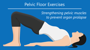
In the early stages of incontinence, the first-line treatment is always pelvic floor physiotherapy and biofeedback - BIOFEEDBACK, by a specialist pelvic floor physiotherapist. Treatment is combined with medication and dietary changes. Conservative treatment with these means brings significant improvement in most cases, but how definitive the treatment will be depends on the underlying problem.
In mild incontinence, neuromodulative therapy for posterior tibial nerve stimulation - POST TIBIAL NERVE STIMULATION - PTNS is applied. Stimulation is done with the help of a small electric stimulator and an electrode that stimulates the posterior tibial nerve (in the leg). Through reflex mechanism, stimulation causes contraction of the pelvic floor muscles. Repeating regular sessions increases pelvic floor tone and improves fecal incontinence as well as urinary incontinence. PTNS is indicated for mild incontinence, whether fecal or urine.
Sphincter injuries during labor are very common and often unavoidable because they depend on the baby's size relative to the size of the perineum, the projection into which the baby descends, and any situations threatening the baby's or mother's safety that may make the completion of labor urgent. Prophylactic episiotomy cannot always protect.
Perineal tears are divided into 4 categories depending on their extent, as shown in the image. Categories 3 & 4 include injuries to the sphincters or the rectum. When tears are noticed immediately and repaired immediately, the problem is usually corrected. Unfortunately, a large percentage of these tears go undiagnosed.
When a sphincter deficit is found due to obstetric or other injury, the issue of chiral repair arises. When the injury is recent, repair is almost always indicated. This is done with a transverse incision in front of the anus, preparation of the internal and external sphincter, overlapping repair, and also reconstruction of the perineal body by folding the lifts to create a supportive surrounding muscle layer.
The surgery yields good results if no nerve damage is present. About 70% of patients see significant improvement in incontinence. However, some will not see improvement and will need to continue other treatments.
If the sphincter deficit developed many years ago and nerve damage has developed, then instead of repair, sacrum-nerve stimulation - SNS may be indicated.
The sacral nerves are stimulated through the implantation of a pacemaker in the sacral part of the spine, which remains as a permanent implant (after a temporary trial). Sacral nerve stimulation - SACRAL NERVE STIMULATION - SNS has very good results in cases of combined urinary and fecal incontinence or in cases where there are sphincter deficits from years of nerve underfunction. Initially, the mature pacemaker is implanted for 2 months, and during this period, it is determined whether improvement is achieved. If there is improvement, the permanent pacemaker is implanted. SNS is now an established treatment with 20 years of experience, and the long-term results are very good, with an effectiveness of around 70%. It has good results in severe constipation, stool incontinence, urinary incontinence, and in many cases pelvic pain. Stimulation of the sacral nerves (SNS) is performed in Athens by a specialist collaborator from our team."
Obstructive defecation syndrome (ODS) is very common, resulting from pelvic muscle relaxation after childbirth or may also be the result of chronic constipation from another cause. ODS is part of pelvic dyssynthesis but has specific radiological findings: first, internal rectal prolapse, and second, rectocele, meaning the rectum protrusion into the vagina.
Obstructive bowel syndrome is diagnosed with bowel proctography or magnetic resonance imaging. Treatment is done with biofeedback, which often yields quite good results, but in more severe cases, surgical treatment is usually needed, meaning correction of the rectocele and the incline-prolapse. Both can be done with surgery, laparoscopic orthopoxy. In this procedure, the rectum is retracted and straightened, eliminating prolapse and rectocele by supporting the rectum to the sacrum. Recovery is rapid and the results are very good.
In half of cases of constipation, pelvic floor dysfunction coexists. Dysfunction causes inability to complete the emptying. This is due to a paradoxical contraction of the heborectal muscle and the sphincters. Contraction without synchronization leads to inability to emptying. Symptoms include a sensation of incomplete emptying, a feeling of blockage, high pressure on the toilet, and the need for finger manipulations to achieve a bowel movement.
The diagnosis is included:
a) with clinical symptoms
b) with proctography/defectomy.
c) with rectal manometry and orthopedic physiology.
Treatment is done through physiotherapy and biofeedback. Surgery has no place.
Chronic pelvic pain is a syndrome with many causes and difficult to treat.
Its various forms are described by names such as:
● Coccygodynia
● Elevator syndrome
● Obstructive neuralgia
● pelvic pain syndrome
● Interstitial cystitis
● Vaginal canal syndrome
Chronic pain often combines three elements: inflammation, muscle spasms, neuropathic pain. Analgesics don't always work. Pelvic floor physiotherapy-biofeedback has some good results.
Some cases respond to infiltration of the lifting muscles and the vulgar nerves, combining muscle relaxants and local anesthetics. In very persistent cases, sacral nerve stimulation (SNS) by implanting a pacemaker in the sacral nerves has been tested with relatively good results. Recent publications suggest that some cases may respond to the new treatment of Functional Magnetic Stimulation (FMS).
This syndrome has recently been described in women. We believe there is congestion of the pelvic veins, especially the left ovarian vein, which causes:
● pelvic pain lasting at least 6 months
● Pain usually on one side (left)
● pain worse with standing
● sudden urgency to urinate
● dilated veins in the genitals, buttocks, thighs
The causes are unclear, but an anatomical abnormality combined with hormonal disorders that cause vasodilation may play a role. Diagnosis is difficult and is made with magnetic angiography. Treatment includes analgesics, hormonal preparations, and, in severe symptoms, embolization of the ovarian veins.
Rectal prolapse is a common condition, but most cases go undiagnosed in the early stages because they begin as internal prolapse. This can gradually develop into external prolapse, which is then noticeable. Rectal prolapse often coexists with a general pelvic floor prolapse and accompanying other pathologies such as rectocele, cystocele, enterocele, etc.
Rectal Prolapse is a common condition caused by a combination of pelvic floor relaxation and increased intra-abdominal pressure. It is much more common in women, where labors relax or even injure the pelvic floor, and pregnancy creates increased intra-abdominal pressure. However, men also suffer from rectal prolapse, either from chronic constipation or from chronic weightlifting (literally, weightlifters are a risk group, as are workers in heavy manual labor). If it exits through the anus, the size of the prolapse matters, as well as whether it is partial or total thickness (that is, whether all the tufts of the rectum or only the mucous membrane protrude).
Symptoms include:
● Pain
● Discomfort
● Constipation
● Difficult rectal emptying
● Fecal incontinence
● Rectal blood
● Mucus from the rectum
If the rectal prolapse is internal and not visible on clinical examination (detected only on radiological examination), it is called an insertion. Recently, the term INTERNAL RECTAL PROLAPSE (INTERNAL RECTAL PROLAPSE) has also been used. Internal rectal prolapse is classified under the OXFORD CLASSIFICATION: GRADE I The prolapse descends as the upper boundary of the rectocele GRADE II It descends to the rectocele but does not reach the GRADE III anal canal It descends to the apex of the GRADE IV anal canal It enters the GRADE V anal canal The prolapse exits the anus and is called EXTERNAL PROLAPSE or TOTAL FAT PROLAPSE
Before treatment, it is important to perform some diagnostic tests to investigate any other coexisting pelvic floor pathologies.
● Colonoscopy (to rule out the possibility of a tumor)
● Magnetic proctography to examine prolapses of other pelvic floor organs such as the rectocele, cystocele, enterocele, which may require simultaneous correction.
● The three-dimensional intrarectal ultrasound to rule out sphincter damage
● Manometry - rectal physiology
Treatment depends on the size of the prolapse and whether it is total thickness.
There are several therapeutic options for rectal prolapse, but the main questions before deciding are: a) Why does the patient have prolapse? If we do not correct the underlying cause, e.g., severe constipation or pelvic relaxation, will problems recur after the surgery? b) If the prolapse is internal (Grade I-IV), has conservative treatment been tested first to improve symptoms? This is necessary before surgery according to the guidelines. c) Is there coexistence of general pelvic floor prolapse with symptoms from the bladder or vagina-uterus? If yes, then cooperation with a urogynecologist is necessary for proper treatment planning.
Pelvic floor physiotherapy and biofeedback also play a role in initial rectal prolapse of the initial stages I-III, which can be applied either as initial treatment to improve symptoms or as complementary therapy to enhance surgical outcome and prevent recurrence. The application of physiotherapy also depends on other coexisting pelvic floor prolapse such as cystocele, etc., and the best option is decided for each patient on a case-by-case basis. Our patients who require such treatments are examined by a pelvic floor physiotherapist to assess the overall treatment plan. Today, it is considered that low-grade prolapse (GRADE I-II) has a good chance of going well with physiotherapy and biofeedback, so we should start from there. High-grade prolapse (GRADE III-IV) does not always respond, and surgery is decided more frequently.
In large total thickness prolapse, the most effective treatment is laparoscopic low rectal resection and simultaneous orthopoxy (Laparoscopic resection-Rectopexy). It is an excellent procedure in terms of treating the problem, but it is a major procedure that is somewhat excessive for elderly patients or individuals with other health issues. For this reason, Laparoscopic Rectopexy is preferred, a high-technique but intermediate seriousness, well tolerated by most patients. It involves laparoscopic preparation of the rectum by separating it from the vagina and adhesions, and then, "coagulation," meaning fixing the rectum to the sacral bone with special sutures and absorbable "pins." The surgery is also described as mesh rectopexy, but in recent years there have been cases where mesh rectopexy causes problems, and although the likelihood of problems is below 5%, I have been using mesh since 2018 and now use exclusively suture and pins (tack rectopexy), which has equally good results. The surgery usually yields excellent results; if there is no previous gynecological procedure, "abdominal orthopoxy" is preferred, which reduces the chances of postoperative constipation. If both rectocele and intestinal hernia are present simultaneously, laparoscopic orthopexy corrects them simultaneously because the entire pelvic floor is lifted and repaired during the procedure.
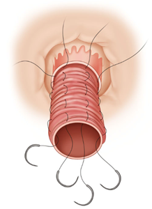
If the prolapse is small and partially thick, then, especially in elderly individuals, corrective surgery is preferred, usually Delorme's surgery, in which the excess mucosa is removed and the rectum folded with sutures. It is an operation without an abdominal incision, and the hospitalization period lasts 1-2 days. Another option is the STARR (Stapled Trans Anal Rectal Resection) procedure, which uses a circular prolapse (Stapler) to extend and remove the prolapse through the rectum (without incision of the abdomen). In the USA, the Altemeier procedure is popular, where the prolapse is completely removed and the final anastomosis is performed.
The ideal surgery for each rectal prolapse may be different for each patient. There are advantages and disadvantages, and to decide whether the patient needs an abdominal or a perineal (transcorrective) method, a study by the specialist surgeon is required, and of course, extensive investigation and discussion with the patient.
In women, rectal prolapse coexists in over 50% of cases with prolapse of other pelvic organs, namely the uterus and bladder, and pathologies called coexistence
● rectocele
● Cystocele
● enterocele
In these cases, it is essential to take the entire pelvis into account, not just the rectum, and to adjust the treatment accordingly. In these cases, cooperation with a specialist urogynecologist is necessary for proper planning of the treatment to be applied.