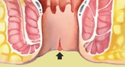
A fissure is a tear of the anal canal. The pain and spasm of the sphincter keep each other together.
They are common and, most of the time, benign. Many people postpone them out of awkwardness or because they fear that every blood is cancer. The test is brief and clarifies the diagnosis.
The anus is a sensitive area of the body affected by a range of pathologies and presents annoying symptoms. Because it is an area that not everyone can easily examine on their own, and it is also not easy for many people to discuss the symptoms of the area with others (even doctors), the result is the creation of significant anxiety. In many cases, anxiety accompanies the symptoms and causes symptoms to worsen and confuse the patient.
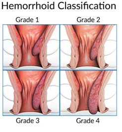
Hemorrhoids are one of the most common clinical problems. They cause annoying symptoms of bleeding, pain, and irritation, and they also cause a lot of anxiety by their presence.
In most cases, they are treated conservatively, but there are also hemorrhoids that definitely require surgery. These are thrombotic hemorrhoids, which present with acute pain and necrosis and can progress to septic conditions. In such cases, emergency surgery is performed. If for any reason this is not possible, they are treated with antibiotics and analgesia.
There are many treatment methods for hemorrhoids; whichever method is followed, the important thing is the proper assessment of the clinical picture and the avoidance of complications.
Hemorrhoids are not a condition. They are a normal network of veins whose purpose is to tightly close the anal area. Like all veins in the body, they stretch when there is high pressure and cause bleeding and other symptoms. Pressure comes from constipation, pregnancy, or sometimes frequent bowel movements in patients with colitis or irritable bowel movements.
A fissure is a tear of the anal canal. The pain and spasm of the sphincter keep each other together.
Symptoms of hemorrhoids include rectal bleeding, pain, burning sensation, itching, prolapse, and discharge.
These symptoms are not unique to hemorrhoids; many rectal and anal conditions appear but have identical symptoms. Because hemorrhoids are very common, they can coexist with any other condition. Diagnosis should be made by a specialist after examining the area. Endoscopy is usually required for blood from the rectum, particularly if the age is over 40.
Hemorrhoids are affected by factors that increase intra-abdominal pressure. Increased intra-abdominal pressure causes blood congestion and swelling of the hemorrhoids. Swelling of hemorrhoids causes symptoms, especially bleeding. The most common factors that increase intra-abdominal pressure are:
a) constipation
b) multiple bowel movements
c) obesity
d) pregnancy
e) sedentary life
g) frequent weightlifting
A diet rich in fiber, fruits, and vegetables is recommended. Fiber supplements are administered. If there is a high frequency of bowel movements, such as irritable bowel syndrome and colitis, these conditions need to be managed to relieve hemorrhoids.
Hemorrhoids often cause problems during pregnancy. It has been found that hemorrhoid thrombosis occurs in about 8% of pregnant women in the last trimester of pregnancy and around 20% after childbirth. Therefore, more than one-third of pregnant women will experience acute and painful hemorrhoid problems. Therefore, prevention is necessary with an appropriate diet, fiber supplements, and mild laxatives during pregnancy. If there are significant symptoms from hemorrhoids and pregnancy is planned, it is better to treat hemorrhoids preventively before pregnancy begins.
The vast majority of hemorrhoids do not require surgery. If these are internal hemorrhoids (1st & 2nd degree), then topical treatments with creams, suppositories, and dietary fiber are sufficient to relieve symptoms. Local care with a lukewarm shower instead of toilet paper and avoiding irritating soaps helps.
When are hemorrhoids operated on? a) When complications occur - thrombosis b) When symptoms—bleeding, pain, irritation, prolapse—do not subside and affect quality of life
There is a wide range of procedures for hemorrhoids, which means none are completely satisfactory. The main goal in hemorrhoid surgery is the absence of pain, since the anus is a very sensitive area to pain. The second goal is to avoid recurrence after surgery. On this page, we will briefly present the main invasive techniques available in European and American guidelines. We will not deal with techniques that are heavily marketed by companies without proof of benefit for the patient, because modern surgery must be based on what is called "Evidence-Based Medicine."
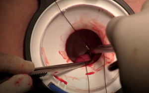
The classic Milligan Morgan hemorrhoidectomy is an extremely effective and safe procedure, especially for grade 4 hemorrhoids. Its drawback is that it has considerable postoperative pain, which is of course controlled with appropriate painkillers. To alleviate this problem, a variation is made with a "closed" Ferguson-type hemorrhoidtomy.
Longo stapler surgery uses a stapler tool to resection the hemorrhoidal ring and simultaneously stapler stitching of the underlying tissues. It is very effective, especially for circulating prolapse hemorrhoids, and has very little pain. It was a popular procedure between 2000-2010 and is still performed today in select cases with excellent results. The problem, however, is that in about 1% of cases, it can cause some serious complications, such as a feeling of urgency to have a bowel movement, and an avaginal fistula. For this reason, it is performed only in selected cases by specially certified surgeons.
The procedure is performed using the LIGASURE device, which uses bipolar electrothrust energy regulated by an electronic brain depending on tissue resistance, resulting in tissue melting and vessel sealing without bleeding and minimal pain. Studies have found LIGASURE surgery to have a signal advantage in terms of pain and healing speed. A similar technique is with the HARMONIC ACE energy source, which uses ultrasound to cut tissues. The LIGASURE and HARMONIC ACE techniques combine the advantages of minor trauma with great effectiveness.
This technique is available under various names, such as HAL -RAR, THD, TRILOGY, etc., which depend on the names companies give their tools. Essentially, it consists of a series of sutures inserted into the hemorrhoids to reduce blood flow. Usually, 6 sutures are inserted, but since these are often not enough to eliminate hemorrhoids, they are supplemented with additional sutures to fold the hemorrhoids inside the anus. To achieve the procedure, a dilator is inserted under general anesthesia into the anus and, through it, an ultrasound probe to detect the arteries. If you click on the paragraph, it shows a video of the procedure from a center where it is performed. The procedure is less painful than the classic Milligan Morgan procedure but involves many more recurrences.
Radio waves are a form of energy during which tissues are changed at a low temperature, around 60 degrees Celsius. In hemorrhoids, this means they can cause thrombosis and simultaneous burns. Radio waves are a relatively new method mainly suitable for stage II-III hemorrhoids. General anesthesia is required. There are no long-term effects regarding recurrence yet.
Laser has achieved great success in dermatology, where its surface radiation is ideal for treating skin lesions. Things are more complicated in hemorrhoids, and so far there is no evidence that it has any advantage. The laser develops a temperature of 600 degrees Celsius, which clots the hemorrhoids, creating a local burn. It can only be applied to internal hemorrhoids, causing them to shrink, but it is unsuitable for external ones because there it will only end in a burn, which obviously will not be painless. If the studies promoted by the companies producing the devices are removed, then the reputable independent studies have shown no advantage of lasers compared to other techniques for hemorrhoids, and for this reason, the laser is not included as a guideline/recommendation in American or British guidelines, etc., which reveals its weak scientific evidence.
A fissure (anal fissure) is a common and painful problem. It is a tear (essentially a traumatic tear) of the skin of the anus that usually occurs due to constipation. In rare cases, there may be another cause, such as Crohn's disease.
The main symptom is severe pain, which may appear during a visit to the bathroom or may be continuous day and night. In some cases, there is also a small amount of blood. A fissure often cannot be detected during the clinical examination because the patient is in so much pain that they cannot tolerate the examination. In such cases, an examination under anesthesia is necessary.
The reason is that the anal area is very sensitive and a strong spasm of the sphincter muscles occurs. This results in reduced blood flow to the fissur, which makes healing difficult.
All treatments aim to resolve the spasm. In earlier years, the practice was sphincterotomy or band dilation. Both of these surgeries had incontinence as a side effect in 5-15% of cases, and for this reason, they are now completely abandoned by specialist rectal surgeons. Treatment today depends on whether the fissure is acute or chronic. A fissure is acute within 8 weeks from the first presentation. From then on, it is defined as years. Treatment is easier at the acute stage.
a) The first treatment is with nitrite ointments. These contain the active substance GTN, which dilates blood vessels and relaxes the sphincter muscles. This increases blood circulation and promotes healing. Treatment is required for 8 weeks, and the success rate is 80%. If there is no healing and symptom improvement, injections are administered. Ointments may cause temporary headaches (due to dilation of the head's blood vessels) as a side effect in some patients; they usually resolve within 2-3 days with a little paracetamol.
b) Injections of muscle-relaxing toxin. They contain the toxin of Clostridium Botulinum, which partially paralyzes the muscles for 2-3 months and similarly improves blood flow and healing. The treatment has a success rate of around 80%.
a) Fissurectomy The term is misleading. What is removed is excess scar tissue that accumulates around the fissure and is considered an additional reason for non-healing. Fissurectomy is combined with repeat injections.
b) Formation of a flap In persistent cases of non-healing fissures, this can be covered by creating a skin flap that covers the fissur, and it heals within 2 weeks. It is a specialized procedure with very good results for difficult cases.
Conclusion: A fissure is a difficult problem that can be treated if a strategy as described above is followed. Under no circumstances should sphincterotomy or ring dilation be performed nowadays, because patients who suffer as a side effect of fecal incontinence will face a more difficult problem. The flap is also necessary after a failed fissure surgery with posterior sphincterotomy that results in a permanent posterior wound with a fistula, known as "keyhole deformity."
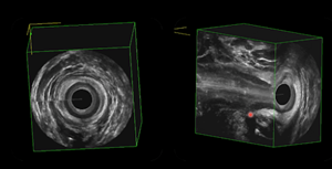
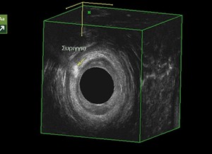
The 3D Intrarectal Ultrasound - 3D EUS is very useful in diagnosing and therapeutic planning of fissures because
a) It depicts not only the superficial fissure but also the deep scarring of the chronic fissure
b) It accurately visualizes the internal and external sphincter, providing great safety in any treatment
c) Diagnoses any sphincter damage from previous treatment or from childbirth
d) Diagnoses any other coexisting pathologies such as fistulas or hemorrhoids, which may cause symptoms confused with fissures
e) provides information on the thickness, anatomy, and condition of the clamps for proper injection application.
Sphincterotomy
Internal collateral sphincterotomy is a treatment that was applied for many years before the discovery of pharmacological treatments with good results in treating fissure pain. Unfortunately, however, it carries an inevitable risk of gas incontinence and even stool, especially in women with a history of childbirth. Although the risk is low, why would a young person risk developing incontinence for life, when currently, over 90% of treatment is possible with medication? As for the remaining percentage, there is always the creation of a flap, which is very effective for chronic stretch marks without causing incontinence. The photo shows the image of the internal sphincter after a sphincterotomy with a large deficit. Many good surgeons still apply it, but personally, I believe that sphincterotomy has closed its historical cycle and should be abandoned.
Peter Lord anal dilation
This method is very aggressive because it involves a large dilation of the anus with four fingers under anesthesia, which simultaneously and circularly breaks the fibers of both the internal and external sphincter. It is naturally effective for fissure pain because the sphincters can no longer have spasms or normal closure. The risk of incontinence with this method is very high, and the results are irreversible. It must be abandoned.
Laser fissure cauterization
Simple cauterization of the fissur, either with laser, diathermy, or whatever else it achieves, is cleaning the scar tissue. So far, this is not a bad thing, but if not combined with some method of relaxing the sphincter—toxin or GTN, it will not have a therapeutic effect and will recur. Another "accident" that sometimes occurs is the application of a laser with excessive zeal, which leads to burning and subsequently rupture of the internal sphincter. Essentially, in these cases, an involuntary sphincterotomy occurs, which, however, takes place at the sixth hour (posterior) and therefore carries a higher risk of incontinence than lateral sphinctertomy.
The periteral fistula is one of the most common and challenging problems in surgical science. It is impressive that even today we apply the classic Hippocrates method extensively, that is, placing a thread inside the fistula (now called a seton) to drain the infection. About 2,300 years later, high-tech methods have not proven more effective!
Essentially, a fistula is an infection that starts from a small abscess, which starts by infecting a small gland in the anus. When the abscess forms, it looks for a way to expel (drain) the pus. This automatically opens a small channel through which pus flows—this is the fistula.
What is the problem? That once the fistula forms, it doesn't close easily. Why? Because new pus accumulates every now and then, which again tries to drain. This creates a vicious cycle of drainage and reinfection that repeats. Why does new pus accumulate? Because the anus contains millions of microbes and cannot be sterilized.
Of course, various factors are involved: local immunity, factors that promote inflammation, diabetes, and more.
Peripheral fistulas are a difficult chronic problem that requires specialization and the right strategy for definitive treatment. St. Mark's Hospital was once founded specifically for the treatment of fistulas.
A fistula is an abnormal communication between two epithelial surfaces. An epithelial is a surface covered by epithelium; the membranes are the skin, the mucous membranes of the mouth, intestine, vagina, bronchi, etc. A peritative fistula (fistula in ano) is an unnatural communication between the skin (the outer opening of the fistula) and either an abscess, the anus, or rectum (the inner opening of the fistula). If the fistula does not have an internal opening, it is called blind and we are not particularly concerned because all blind fistulas close on their own relatively quickly. The problem lies in the presence of the inner opening. Fistulas are classified according to their relationship to the sphincters, which determines both the choice of treatment and the prognosis. Superficial fistulas are relatively easy to treat, but generally, the greater the involvement of the sphincters in the fistula, the more complex the treatment.
There is a close relationship between the perianal abscess and the fistula. The abscess is always present, and the fistula is the mechanism by which the human body tries to heal the abscess. So wherever there is a fistula, there is or has been an abscess. Every perianal abscess must be drained as quickly as possible unless it disappears completely with antibiotics. Otherwise, it will develop into a fistula. That is why it is important that when a perital abscess appears, it is treated promptly and with full drainage; otherwise, it is often followed by a fistula. If the fistula creates only an external opening (on the skin), the patient is lucky and will close on its own soon. However, if drainage happens simultaneously with an internal opening in the rectum, things become difficult.
There are 2 difficulties that cause fistulas to recur.
a. The anus cannot be sterilized—so infected material continuously flows into the cavity of the previous abscess, causing chronic infection.
b. Fistulas are in close contact with the sphincters and therefore cannot always be surgically removed decisively (there is a risk of incontinence).
The first symptoms come from the periteral abscess and include local swelling, severe pain, and fever. When the abscess drains through fistula formation, these symptoms gradually subside and are replaced by discharge, which, depending on the phase, may be pus, fecal material, bloody, or yellow serous.
The symptoms of the fistula have a cyclical course. After the initial drainage, the symptoms subside within a few days, leaving only fluid secretion, which gradually clears up. The patient believes they have been cured, especially if, in many cases, the external opening is completely closed. Unfortunately, the calm usually lasts only a few weeks or months, at which point the symptoms return even stronger. The reason is that the internal opening of the fistula remains open, which is not visible and continues to supply the cavity with soaked material from the intestine. A new abscess forms, which requires new drainage. The symptoms then subside again until further recurrence. Relapses should be avoided because they can lead to increasingly complex fistulas and there is a small risk of developing perineal gangrene, which is life-threatening.
The most widespread radiological method is magnetic resonance imaging, which shows the fistula's course and anatomical relationships. Another modern method is three-dimensional intrarectal ultrasound - 3D Endorectal Ultrasound (3D EUS), which allows three-dimensional imaging of the fistula and also allows for the determination of whether the duct is open. Ultrasound has advantages - it can be repeated many times during monitoring at low cost, painlessly, and without the patient's inconvenience.
The principles of treating fistulas are ancient and have not been changed by any new method.
Principle one – drainage of sepsis (i.e., pus). This is not only necessary during surgery but also continues postoperatively, e.g., with seton placement.
Second principle – removal of the fistula duct. Ablation can be done in many ways: removal of the fistula, cauterization with diathermy, radio waves, laser, etc. Also, removal of the duct is done by filling the fistilla with materials. Removal of the fistula should ONLY AFTER COMPLETE DRAINAGE AND TREATMENT OF THE SEPTA!
Principle three – closing the internal opening of the fistula (to stop continuous infection from the intestine).
Fourth principle – all surgical procedures must be performed with care and respect for maintaining the integrity of the clamps.
Whatever method is followed for treatment, regardless of technique, special equipment, etc., the principles of treatment remain the same and must be respected for success.
There is a wealth of surgical techniques for treating fistulas. The techniques being developed are trying to become increasingly less invasive and painless. Some techniques have advantages for specific types of fistulas. All techniques have the same long-term effectiveness, about 70% success. This means that no matter which method is used, there is always a recurrence rate of 30%. Recurrences are due to recurrence of infection (which depends on many factors). This must be clear to the patient from the start to avoid disappointment and distrust.
Complete resection of the fistula—"fistrectomy"—is only possible if it leaves the external sphincter intact and does not have an internal opening. In some cases, in men, it is possible to safely dispense a small part of the external sphincter without problem, but it is generally preferable to avoid it. Complete "fistectomy" is generally difficult to achieve often. A substitute is partial removal up to the edge of the sphincter, called "fisticide."
The seton – seton – is an ancient method, described by Hippocrates, which controls the vicious cycle of sepsis—abscess—drainage through the fistula. The seton is either a suture (nauloon thread) or an elastic thread that keeps the fistula's duct open and prevents infection from recurring. The seton is inserted into the fistula and exits through both the inner and outer openings; it is painless and remains in place for several months. How long it lasts depends on the course of the decay.
There are 2 types of setons. The Hippocrates or "tight" seton, which is a nylon thread that gradually passes through the clamp like a wire through ice and removes the fistula.
The "loose" seton, which acts as a drain of pus and remains for many weeks until the infection clears up.
An endoscopic method that requires specialized equipment and experience. We are already successfully applying this method in the Department of Colon-Rectal Surgery at Metropolitan General. The method uses an endoscopic "Fistuloscope" which enters through the external opening of the fistula and examines the fistula's duct from the inside, discovering any additional pores and abscesses. At the same time, the duct is washed and endoscopically the internal opening of the fistula is discovered. The scar and infected tissue is cauterized and endoscopically removed with special micro-instruments without the need for large incisions or wounds. After complete cleaning and debridement of the infected tissue, the internal opening is closed with flap creation. The outer mouth remains open for drainage. The advantages of the method are:
– the formation of large surgical wounds is avoided; there is only a small hole in the outer opening of the fistula
– all hidden fistula ducts and abscess are found and cleaned from the inside
– The internal opening of the fistula, which is responsible for relapses, is closed
– no hospitalization is required and recovery is very fast
The VAAFT method is mainly suitable for long and blind fistulas, the so-called euthyschiatic ones.
LIFT surgery is a variation of the classic fistula-incision. The fistula is carefully separated from the sphincter and its two ends are destroyed and ligated separately.
Treatment with flap creation is the most effective fistula treatment. In it, a tissue flap is created either from intestinal mucosa or a skin flap, which is transported and covers the internal opening of the fistula. It is a larger procedure than the others, but because it has very good results in the hands of specialists, it is suitable for difficult, chronic fistulas in which other treatments have failed.
The use of a laser always sounds appealing and non-invasive, and to some extent, it is so in many medical specialties. In perioral fistula surgery, the laser can be used instead of electrical diathermy for better energy distribution during debridement, but it is not clear from the literature whether it has an advantage. It is used for cauterizing the fistula duct and is an easy technique that can be done without general anesthesia, but the success of fistulastic treatment is not based solely on the laser but on overall adherence to the principles mentioned above and on the surgeon's experience, which is the most important. Therefore, if there is no cost issue (the laser adds about 500 euros to the cost of the operation), then the laser can offer a useful complement to surgical techniques, but it cannot provide treatment on its own.
Filling the fistulle with biological materials has been a common practice for twenty years. Various materials have been tested, such as fiber glue, a collagen plug, collagen paste, etc. The method is successful but only if combined with another surgical procedure, such as closing the inner opening. This has been tested in various ways, either simple staple with biological material or by creating a flap. The latest development is the use of an organic Cyanyl-Acrylate adhesive, which has a higher chance of success.
Treatment of fistulas is characterized by many recurrences. No method has a clear advantage for all cases. All methods, after initial excitement, prove to be about 70% effective in the long term, meaning about 30% recur. This means reoperation. That is why it is important that surgeries are conservative and with as little trauma as possible; this is the purpose of all new techniques. However, when failures and recurrences have occurred, then a flap operation (mucosal advancement flap) should be performed by an experienced rectal surgeon, which provides the best definitive results. But all methods have their place.
When patients ask, "Which method do you use?" the answer is, "All of them! Each method for the fistula where it has an indication and better chances of success."
There is always the patient's concern that surgery on a fistula will cause incontinence. Only the procedure is guaranteed by certified bowel and rectal surgeons specializing in fistulas. The risk of the sphincter being cut during the operation is very small to negligible in the hands of a trained surgeon. On the other hand, any incomplete and incomplete procedures that allow prolonged septic status can cause incontinence due to inflammation. Chronic inflammation of the sphincter causes fibrosis, impaired function, and incontinence.
Intrarectal ultrasound is a very useful tool not only for initial assessment/diagnosis but also for easy, frequent monitoring of the postoperative fistula course and prevention of recurrence. It is a simple, harmless, fast, and painless examination that provides valuable information about the course of sphincter infection and pus collection. At the Bowel - Rectal Clinic, a 3D intra-rectal hyperechography is performed on all patients with fistulas to determine the optimal treatment.
Fistulas between the rectum and other organs are common. Orthocystic and rectovaginal fistulas are primarily caused by diverticulitis, but they can also be caused by tumors, Crohn's disease, radiation therapy, and others.
Orthovaginal fistulas also occur as a complication of obstetric sphincter injury and pose a major public health problem in some third-world countries.
Treatment is surgical. The prognosis is good when the operation is successful.
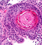
It is a common condition in young people and especially in hairy men. It has been suggested to have a congenital cause, but today most believe it simply occurs from hair entering the skin in men with a deep gluteal cleft. It gives annoying symptoms of an infection-abscess, but surgical treatment should be done carefully and, if possible, minimally invasive, so that the treatment does not end up more bothersome than the problem!
Coccyx cyst is a common condition that usually affects young people. There is disagreement about the causes, but the prevailing view is that hairs infiltrate the skin that form an abscess. Hairy men have a much higher prevalence of the condition.
The coccyx cyst manifests as pain, redness, and swelling in the slit between the buttocks (natal cleft). Symptoms worsen and end in an abscess that either breaks on its own or is operated on and drained. Some cases resolve when antibiotics are taken immediately.
Treatment of a coccyx cyst is initially "easy," but the problem is recurrences. If antibiotics are taken without surgery, symptoms subside for weeks or months, but there is a high rate of recurrence. The same happens if there is a simple opening/drainage. The best treatment method is surgery when the cyst does not have an acute infection. Simple removal by suturing the wound (traditional "closed" method) is the worst option because it often results in wound fragmentation and delayed healing (weeks or months). To prevent this, the "open" method is also applied, where the wound is simply left open to heal for a second purpose. However, it takes a lot of time, which is why it is not much better than the closed method. Because these healing problems exist—the wounds of the midline between the buttocks experience constant tension due to movement, sitting, etc. So, to avoid healing problems, the procedure must have one goal. Removal of the cyst without trauma to the midline.
The destruction of the coccygeal fistula of the bladder follows the path of new treatments for perianal fistulas. This is done using the VAAFT syringoscope. With the syringoscope, the entire cyst can be nucleated and all the hairs causing recurrence removed without any trauma. The results are usually very good and recovery is very rapid. The procedure is called EPSiT = Endoscopic Pilonidal Sinus Treatment.
A similar procedure can be performed with a LASER fiber, in which case the procedure is called SILaC .
If the cyst is large, complicated, a cyst recurrence, or with multiple fistulas, it is removed with wide excision and then the wound is closed with a plastic flap that is far from the midline. The most common flap is the Karydakis flap. The flap surgery yields excellent results.
As with perierative fistulas, with coccyx cyst fistulas, the nucleation/destruction of the fistula can be aided by the use of biological glue. The fistula can be nucleated using the EPSiT method or with LASER, and then the duct can be filled with biological adhesive. The adhesive can be repeated in a second session. A recent study from Britain showed effectiveness in over 90% of cases with this technique.

Anal cancer is a rare disease and mainly affects people over 60 years old. Predisposing factors include:
● HPV virus (warts)
● HIV virus
● chronic inflammation (fistulas, Crohn's disease)
● immunosuppression
The symptoms are not specific: pain, blood, tenesmus, masses of inguinal lymph nodes. Sometimes anal cancer is found randomly in an excluded hemorrhoid.
Treatment depends on the stage at diagnosis. At an early stage, up to the submucosa, local excision may be sufficient. However, when there is deeper infiltration, chemotherapy and radiotherapy are used as the first line. The results are good in the majority of cases. When recurrence occurs, surgery is performed—abdominal peripineal excision with colostomy.
Pain in the anus is a very common symptom. Anxiety and stress almost always coexist, but the label of a "psychological" problem should not be used, because, first, there is almost always an "organic" cause, and second, because every psychological cause causes physical problems in the gastrointestinal tract.
Anal pain can take various forms: sharp pain, a burning sensation, tensure, inability to sit or walk, etc. The location of the pain may shift or be difficult to pinpoint.
The diversity of features is due to the fact that all pelvic innervation is supplied by the sacral nerves, which causes diffusion of the sensation of pain and other sensory disturbances.
The forms of pain can be:
● Coccygodynia
● Proctodynia - Proctalgia Fugax
● Elevator Syndrome
Treatment is mainly biofeedback and pharmacology. Muscle-relaxing injection of the lifts with botox, combined with a block of the obedient nerves, may have relieving effects.
One particular form may be Alcock's Channel Syndrome, in which the vulgar nerve can become trapped and sharp pain can occur. Pain is worse when sitting, and patients prefer to stand upright. A burning sensation and a foreign body sensation in the anus may coexist. Treatment involves infiltration of the Alcock channel, combining steroids and local anesthetics. Surgical release of the nerve has also been described in rare cases.
